ECG
- Home
- / ECG
Explore and Review ECG
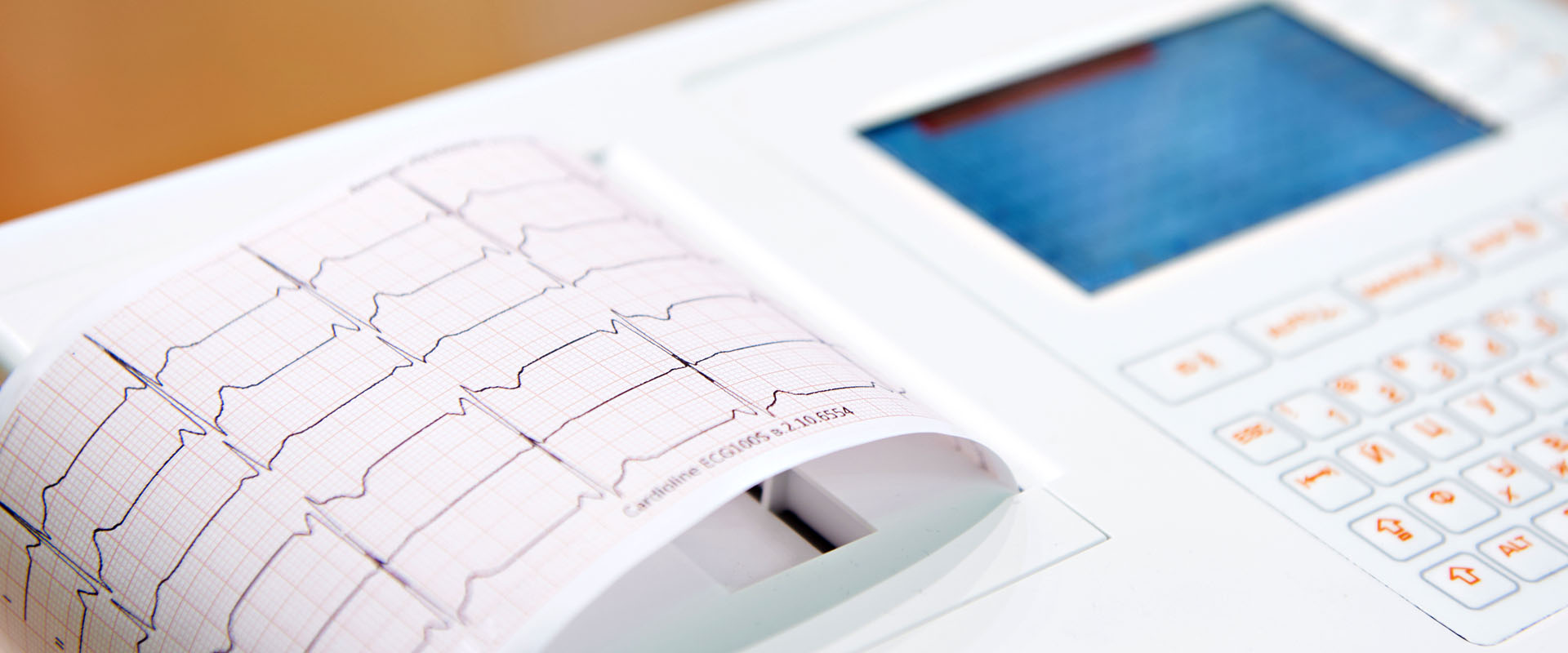
Electrocardiography (ECG or EKG) is a vital diagnostic tool used to assess the electrical activity of the heart. This non-invasive procedure records the heart’s rhythm and can reveal various cardiac conditions. In this comprehensive exploration, we delve into the intricacies of ECG, shedding light on different rhythms and their significance.

Sinus Rhythm - Heart Rate 72
Normal Sinus Rhythm (NSR), also referred to as Regular Sinus Rhythm (RSR), stands as the prevalent adult cardiac rhythm, typically maintaining rates within the range of 60 to 100 beats per minute. This rhythm manifests with narrow QRS complexes and upright P waves, particularly prominent in Lead II.
Sinus Bradycardia – Heart Rate 54
Sinus Bradycardia is characterized by a heart rate less than 60 beats per minute, and in this instance, it manifests at 54 beats per minute. While this may be well-tolerated by healthy individuals, it can also occur in athletes due to optimal cardiac stroke volume, requiring fewer heartbeats to maintain effective cardiac output. Additionally, Sinus Bradycardia might result from Vagal stimulation or be associated with Sick Sinus Syndrome. Recognized by a narrow QRS complex and upright P waves in Lead II, understanding this rhythm variation is crucial for healthcare professionals and EKG technicians in providing comprehensive patient care.
Sinus Tachycardia – Heart Rate 138
Sinus Tachycardia, clocking in at 138 beats per minute, offers a glimpse into the accelerated rhythm of the heart. Characterized by a narrow QRS complex and upright P waves in Lead II, this phenomenon provides a factual perspective for students studying EKG interpretation. Understanding the intricacies of Sinus Tachycardia at a heart rate of 138 BPM contributes to the foundational knowledge essential for students in the field of electrocardiography.
Sinus Arrhythmia – Heart Rate 78
Sinus Arrhythmia at a heart rate of 78 signifies an irregularity in the pace of the heart’s natural rhythm. While maintaining a steady average of 78 beats per minute, this arrhythmia introduces variations in the time interval between heartbeats. Identified by a narrow QRS complex and upright P waves in Lead II, this condition highlights the nuanced nature of sinus rhythm irregularities. Studying Sinus Arrhythmia at this heart rate provides a practical understanding of how the heart’s rhythm can exhibit subtle yet distinctive variations within a specific range.
Sinus Exit Block – Heart Rate 48
A Sinus Exit Block at a heart rate of 48 indicates an interruption in the electrical signals as they leave the sinus node, temporarily disrupting the heart’s natural pacing. This phenomenon is characterized by a sudden pause in the heart’s electrical activity, leading to a temporary halt in heartbeat generation. Observing this condition with a heart rate of 48 allows students to explore the dynamics of electrical conduction within the sinus node and the challenges associated with these brief interruptions. Understanding Sinus Exit Block at different heart rates contributes to a comprehensive grasp of cardiac rhythm irregularities.
Sinus Arrest – Heart Rate 54
At a heart rate of 54, Sinus Arrest signifies a temporary cessation of electrical impulses originating from the sinus node. This cardiac phenomenon leads to a pause in the heart’s rhythm, creating a brief interruption in the regular heartbeat sequence. Students exploring Sinus Arrest at this heart rate gain insights into the intricacies of electrical signal interruptions within the sinus node, comprehending the transient nature of these pauses and their impact on overall cardiac rhythm. Delving into Sinus Arrest at a heart rate of 54 offers a nuanced perspective on temporary halts within the sinus rhythm.
NSR with Pac - Heart Rate 84
Premature Atrial Complexes or PAC result from irritability to the atria resulting in increased automaticity of atrial tissue. Since the atria initiate an impulse earlier than expected from the SA node, this is a premature complex. Expect narrow QRS and flattenned, notched, peaked or biphasic P waves for the PAC.
SVT – Heart Rate 180
At a heart rate of 180, Supraventricular Tachycardia (SVT) unfolds as a rapid and sustained heartbeat originating above the heart’s ventricles. Students exploring SVT at this heart rate delve into the intricacies of abnormal electrical pathways, leading to swift and often palpable palpitations. Understanding SVT at 180 beats per minute equips learners with insights into the challenges posed by abnormal conduction, shedding light on the mechanisms that contribute to this particular cardiac arrhythmia.
Atrial Fibrillation – Heart Rate 90
A heart rate of 90 in Atrial Fibrillation marks a state of irregular and often rapid heartbeats originating in the atria. In this condition, the atria lose their coordinated rhythm, leading to chaotic electrical signals. This description elucidates the specific manifestation of Atrial Fibrillation at a heart rate of 90 beats per minute, providing a glimpse into the disarrayed atrial activity characterizing this cardiac arrhythmia.
Atrial Flutter – Heart Rate 75
A heart rate of 75 in Atrial Flutter signifies a distinctive cardiac rhythm characterized by rapid, regular contractions of the atria. In this condition, the heart’s upper chambers beat in a unique, organized pattern, often creating a “fluttering” sensation. This description encapsulates the specific manifestation of Atrial Flutter at a heart rate of 75 beats per minute, offering insight into the orchestrated atrial activity that defines this particular arrhythmia.
Paced Atrial – Heart Rate 60
A heart rate of 60 in Paced Atrial rhythm reflects a scenario where the heart’s natural pacing mechanism is supplemented or replaced by artificial pacing. This controlled pacing, often initiated by a pacemaker, orchestrates the atrial contractions at a steady rate of 60 beats per minute. In this context, the description unveils a deliberate intervention to regulate heartbeats, emphasizing the precision and guidance provided by the pacemaker in maintaining a heart rate of 60.

NSR with 1 AVB - Heart Rate 74
Premature Atrial Complexes (PAC) occur due to heightened irritability in the atria, leading to increased automaticity of atrial tissue. As the atria generate an impulse earlier than anticipated from the SA node, it results in a premature complex. Characteristics include narrow QRS complexes and P waves that are flattened, notched, peaked, or biphasic for the PAC.
2 AVB Type 1 – Heart Rate 48
A heart rate of 48 in Second-Degree Atrioventricular Block (Type 1) signifies a distinctive cardiac conduction pattern. This rhythm reflects intermittent delays in the electrical signals traveling from the atria to the ventricles, leading to occasional dropped beats. The deliberate slowing down of the heart’s conduction system unfolds a narrative of nuanced navigation within the atrioventricular pathways, where the communication between the heart’s chambers follows a unique and rhythmically deliberate course.
2 AVB Type 2 – Heart Rate 60
With a heart rate of 60, Second-Degree Atrioventricular Block (Type 2) unfolds a complex tale of cardiac conduction. This rhythm showcases intermittent disruptions in the electrical communication between the atria and ventricles. Each beat becomes a deliberate orchestration, revealing the intricacies of an AV block where some atrial impulses fail to reach the ventricles. The steady cadence of 60 beats per minute underscores the meticulous dance between the heart’s upper and lower chambers, highlighting the unique characteristics of Atrioventricular Block Type 2.
2 AVB 2:1 – Heart Rate 38
At a heart rate of 38, the rhythm of Second-Degree Atrioventricular Block (Type 2) with a 2:1 conduction ratio unfolds a distinctive cardiac symphony. In this intricate composition, every other atrial impulse manages to traverse the atrioventricular node, reaching the ventricles in a synchronized manner. The deliberate pace of 38 beats per minute unveils a captivating dance, where the heart’s communication experiences a rhythmic ebb and flow, creating a unique pattern within the realm of atrioventricular blocks. Explore the harmonious yet intricate nature of 2:1 AV block at a heart rate of 38
3 AV Block – Heart Rate 36
With a heart rate of 36, the cardiac symphony takes on a complex and deliberate cadence, signaling the presence of Third-Degree Atrioventricular Block. In this intricate composition, the atria and ventricles dance to a distinct rhythm, each moving independently. The absence of synchronicity between atrial and ventricular contractions creates a unique pattern, where the heart’s communication undergoes a profound disruption. At 36 beats per minute, delve into the intricacies of Third-Degree AV Block, where the orchestra of the heart plays a composition marked by independence and separation.
NSR with PJC - Heart Rate 84
Premature Atrial Complexes (PAC) occur due to heightened irritability in the atria, leading to increased automaticity of atrial tissue. As the atria generate an impulse earlier than anticipated from the SA node, it results in a premature complex. Characteristics include narrow QRS complexes and P waves that are flattened, notched, peaked, or biphasic for the PAC.
Junctional Rhythm – Heart Rate 48
At a heartbeat cadence of 48, Junctional Rhythm orchestrates a unique musical arrangement within the heart’s chambers. In this specialized rhythm, the electrical impulses emanate from the atrioventricular (AV) junction, steering the cardiac performance. With an intentional pace of 48 beats per minute, Junctional Rhythm takes the lead, shaping a distinct harmony that differs from the regular sinus rhythm. Dive into the world of Junctional Rhythm at 48 beats per minute, where the AV junction assumes the conductor’s role, directing the cardiac symphony with precision and purpose.
Accelerated Junctional – Heart Rate 82
Accelerated Junctional Rhythm with a heart rate of 82 signifies an increased pacing of heartbeats originating from the atrioventricular (AV) junction. In this rhythm, the AV junction takes over the role of the heart’s pacemaker, resulting in a faster heart rate of 82 beats per minute. This accelerated rhythm is a notable aspect of the heart’s electrical activity and can be identified through an electrocardiogram (ECG). Understanding this cardiac pattern is crucial for healthcare professionals involved in EKG monitoring and interpretation.
Junctional Tachycardia – Heart Rate 186
Junctional Tachycardia with a heart rate of 186 indicates a swift and heightened heartbeat originating from the atrioventricular (AV) junction. In this tachycardic rhythm, the AV junction takes charge of the heart’s pacing, resulting in a rapid heart rate of 186 beats per minute. This distinctive cardiac pattern is recognizable through an electrocardiogram (ECG) and holds significance in the realm of EKG monitoring and interpretation. Healthcare professionals and students in this field benefit from understanding the characteristics and implications of Junctional Tachycardia
Wandering Pacemaker – Heart Rate 78
Wandering Pacemaker, characterized by a heart rate of 78 beats per minute, refers to a dynamic atrial pacing pattern. In this cardiac rhythm, the pacing impulse shifts between the sinoatrial (SA) node and adjacent atrial tissue, leading to variations in the heart rate. The heart’s natural pacemaker, the SA node, alternates with neighboring atrial sites, resulting in a slightly irregular rhythm. This phenomenon, observed at a heart rate of 78, is an intriguing aspect of EKG interpretation and plays a role in the broader spectrum of atrial rhythms. Understanding the dynamics of Wandering Pacemaker is valuable for individuals engaged in EKG monitoring and interpretation, providing insights into the complexities of cardiac electrical activity.
NSR with PVC - Heart Rate 68
Within the context of a sinus rhythm, the emergence of premature ventricular complexes (PVCs) often signals increased automaticity in the ventricles or the possibility of a reentry phenomenon. While PVCs can be benign, their presence may also indicate irritability in the ventricles, necessitating careful consideration. Recognized by their early arrival in the cardiac cycle, PVCs are characterized by a wider QRS complex, typically exceeding 0.12 seconds. Notably, the T wave associated with PVCs tends to point in the opposite direction from the regular QRS complex. Arranging in distinctive patterns, such as ventricular bigeminy (PVC occurring every second complex) or ventricular trigeminy (PVC every third complex), these variations add a unique dynamic to the cardiac rhythm.
Adioventricular – Heart Rate 38
Adioventricular rhythm, denoted by a heart rate of 38 beats per minute, signifies a distinctive atrioventricular (AV) conduction pattern. This rhythm involves a slower-than-normal passage of electrical signals from the atria to the ventricles, leading to a reduced heart rate. With a heart rate of 38, this condition falls within the bradycardia range.
In Adioventricular rhythm, the atria and ventricles operate independently to some extent, contributing to a lower and often irregular heart rate. This phenomenon may be associated with certain cardiac conditions or conduction system abnormalities. Clinically, recognizing and understanding Adioventricular rhythm is crucial for healthcare professionals, especially those involved in EKG interpretation and monitoring, as it sheds light on the complexities of AV conduction disorders.
Accelerated IVR – Heart Rate 84
A rhythm characterized by inherent ventricular activity at a heart rate of 84 beats per minute, reflecting independent impulses originating in the ventricles. Often transient, it can occur in certain clinical contexts, providing insights into ventricular electrical dynamics.
VTach – Heart Rate 210
Ventricular tachycardia is a rapid, regular beating of the heart originating in the ventricles. With a heart rate of 210 beats per minute, it signifies a potentially serious arrhythmia that warrants medical attention. VTach may compromise cardiac function and necessitate intervention to restore normal rhythm.
VFib – Heart Rate 0
A chaotic and rapid quivering of the heart muscles in the ventricles, leading to an ineffective pumping of blood. In VFib, the heart rate is essentially chaotic and there is no discernible heartbeat. It is a medical emergency that requires immediate intervention such as defibrillation.
Paced Ventricular – Heart Rate 80
Refers to a cardiac rhythm where the heartbeat is controlled or paced by an artificial pacemaker located in the ventricles of the heart. In this scenario, the heart rate is set by the pacemaker, ensuring a consistent and controlled rhythm. The heart rate for a paced ventricular rhythm is measured at 80 beats per minute.
As we conclude our exploration of ECG rhythms, we recognize the intricate symphony that orchestrates the beating of the heart. This page serves not only as an informative guide but also as a testament to the complexities that EKG technicians navigate daily. For those embarking on a journey into the world of EKG monitoring, our comprehensive courses are tailored to equip you with the skills and knowledge needed to decipher these rhythmic patterns. At EKG Monitor Tech in Van Nuys, California, we are dedicated to providing top-notch education, empowering aspiring EKG technicians to excel in their profession. Join us in unraveling the mysteries of ECG, and let the heartbeat of healthcare become second nature to you.


